
Forest
Beloved Grand Eagle
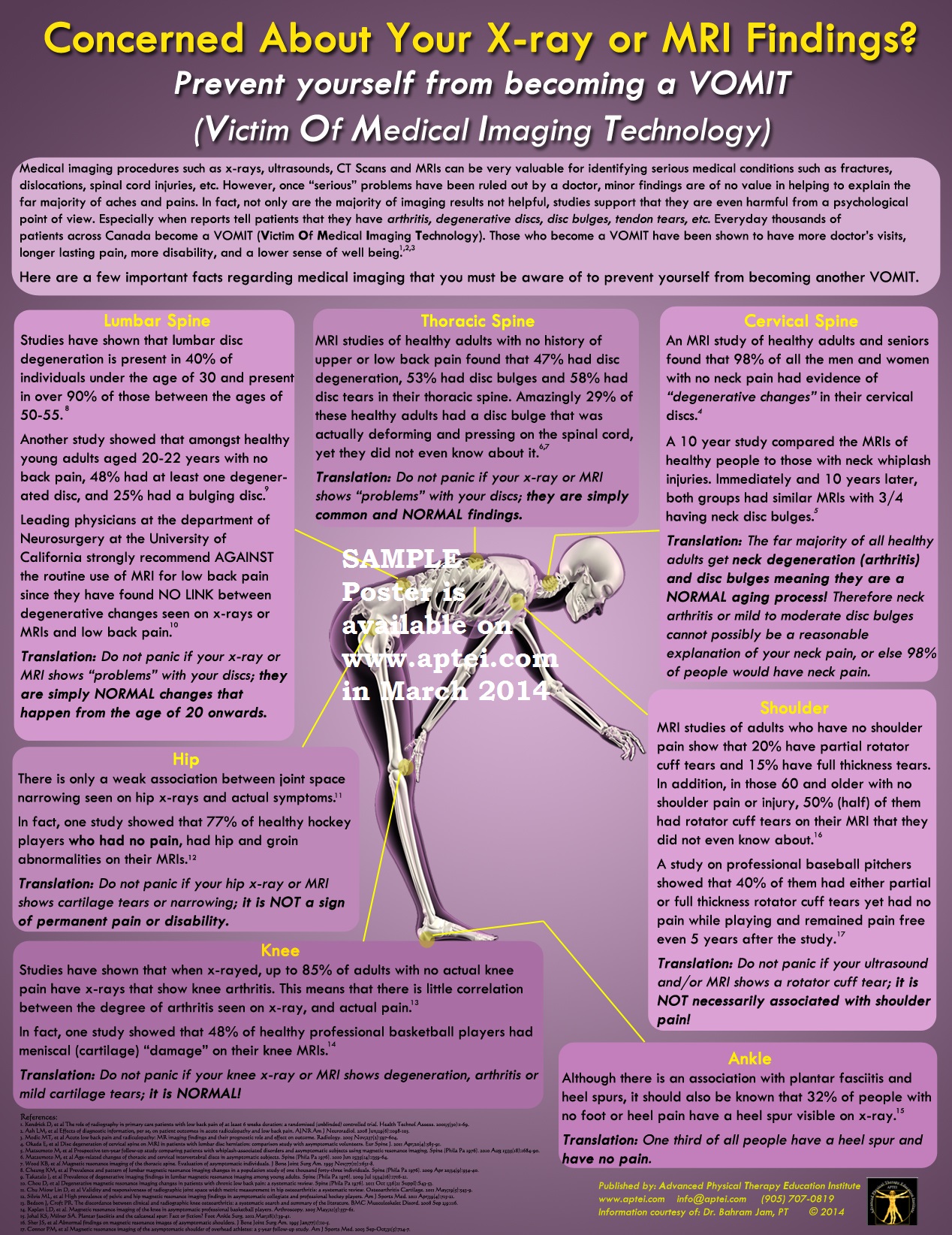
The poster is created by the Advanced Physical Therapy Education Institute. It's great to see physical therapists coming to understand the consequences of the incontrovertible medical evidence: that our bodies are strong and that medical imaging can have negative effects on people.
Here's a direct link to the image:
http://www.aptei.com/shop/index.php?main_page=popup_image&pID=43
The poster is meant to be put up in doctors offices and Physical Therapy offices. You can order it here:
http://www.aptei.com/shop/index.php?main_page=product_info&cPath=0&products_id=43
Of course, it's always important to see a properly licensed medical practitioner to rule out anything serious such as a fracture, cancer, or dislocation.
Here's some information about the poster from the publisher:
VOMIT (Victim of Medical Imaging Technology)
There has been an overwhelmingly positive response to the information on this poster. Please have the small card version of the poster at hand to show your patients who have been told they have DDD, disc bulges, arthritis and rotator cuff tears.
I urge you to hang up the full size VOMIT poster at your clinic and get one as a gift for a referring physician (see www.aptei.com/shop). I have yet to have an MD who disagreed with the information on the poster. There are two reasons why MDs do not typically disclose the information on the VOMIT poster:
1. They simply do not have time to give a full explanation of their patient's radiological report. It is just easier to say, "Yes, your neck x-ray shows you have degeneration at multiple levels"
2. They truly are not aware of the research studies on normal prevalence and epidemiology. MDs genuinely do not know that 75% of asymptomatic people have cervical disc bulges, 85% of asymptomatic adults have knee OA, etc. so when MDs see the radiological reports, they panic which makes the patients panic even more.
If only all MDs knew about and shared the information on the VOMIT poster, I truly believe it would save the Canadian health care system millions of dollars.
I just don't know how we can spread the information to MDs other than one at a time!
VOMIT: Lumbar Spine
Reference:
1. Cheung KM, et al Prevalence and pattern of lumbar magnetic resonance imaging changes in a population study of one thousand forty-three individuals. Spine (Phila Pa 1976). 2009 Apr 20;34(9):934-40.
2. Takatalo J, et al Prevalence of degenerative imaging findings in lumbar magnetic resonance imaging among young adults. Spine (Phila Pa 1976). 2009 Jul 15;34(16):1716-21.
3. Chou D, et al Degenerative magnetic resonance imaging changes in patients with chronic low back pain: a systematic review. Spine (Phila Pa 1976). 2011 Oct 1;36(21 Suppl):S43-53.
Studies have shown that lumbar disc degeneration is present in 40% of individuals under the age of 30 and present in over 90% of those between the ages of 50-55.(1)
Another study showed that among healthy young adults aged 20-22 years with no back pain, 48% had at least one degenerated disc, and 25% had a bulging disc.(2)
Leading Physicians at the department of Neurosurgery at the University of California strongly recommend AGAINST the routine use of MRI for low back pain since they found NO LINK between degenerative changes seen on x-rays or MRIs and low back pain.(3)
Translation: Do not panic if your x-ray or MRI shows "problems" with your discs, they are simply NORMAL changes that happen from the age of 20 on wards.
Reference:1. Matsumoto M, et al Age-related changes of thoracic and cervical intervertebral discs in asymptomatic subjects. Spine (Phila Pa 1976). 2010 Jun 15;35(14):1359-64.
2. Wood KB, et al Magnetic resonance imaging of the thoracic spine. Evaluation of asymptomatic individuals. J Bone Joint Surg Am. 1995 Nov;77(11):1631-8.
An MRI studies of healthy adults with no history of upper or low back pain found that 47% had disc degeneration, 53% had disc bulges and 58% had disc tears in their thoracic spine.
Amazingly 29% of these healthy adults had a disc bulge that was actually deforming and pressing on the spinal cord, yet they did not even know about it.(1,2)
Translation: Do not panic if your x-ray or MRI shows "problems" with your discs, they are simply common and NORMAL findings.
VOMIT: Cervical Spine
Reference:1. Okada E, et al Disc degeneration of cervical spine on MRI in patients with lumbar disc herniation: comparison study with asymptomatic volunteers. Eur Spine J. 2011 Apr;20(4):585-91.
2. Matsumoto M, et al Prospective ten-year follow-up study comparing patients with whiplash-associated disorders and asymptomatic subjects using magnetic resonance imaging. Spine (Phila Pa 1976). 2010 Aug 15;35(18):1684-90.
An MRI study of healthy adults and seniors found that 98% of all the men and women with no neck pain had evidence of "degenerative changes" in their cervical discs.(1)
A 10 year study of compared the MRIs of healthy people to those with neck whiplash injuries. Basically immediately and 10 years later both group had similar MRIs with 3/4 having neck disc bulges.(2)
Translation: The far majority of all healthy adults get neck degeneration (arthritis) and disc bulges meaning they are a NORMAL aging process! Therefore neck arthritis or mild to moderate disc bulges cannot possibly be a reasonable explanation of your neck pain, or else 98% of people would have neck pain.
VOMIT: Hip
Reference:1. Chu Miow Lin D, et al Validity and responsiveness of radiographic joint space width metric measurement in hip osteoarthritis: a systematic review. Osteoarthritis Cartilage. 2011 May;19(5):543-9.
2. Silvis ML, et al High prevalence of pelvic and hip magnetic resonance imaging findings in asymptomatic collegiate and professional hockey players. Am J Sports Med. 2011 Apr;39(4):715-21.
There is only a weak association between joint space narrowing seen on hip x-rays and actual symptoms.(1)
In fact one study showed that 77% of healthy hockey players who had no pain, had hip and groin abnormalities on their MRIs.(2)
Translation: Do not panic if your hip x-ray or MRI shows cartilage tears or narrowing, it is NOT a sign of permanent pain or disability.
VOMIT: Shoulder
Reference:1. Sher JS, et al Abnormal findings on magnetic resonance images of asymptomatic shoulders. J Bone Joint Surg Am. 1995 Jan;77(1):10-5. 2. Connor PM, et al Magnetic resonance imaging of the asymptomatic shoulder of overhead athletes: a 5-year follow-up study. Am J Sports Med. 2003 Sep-Oct;31(5):724-7.
MRI studies of adults who have no shoulder pain show that 20% have partial rotator cuff tears and 15% have full thickness tears. In addition, in those over the age of 60, 50% (half) of those who had no shoulder pain or injury had rotator cuff tears on their MRI that they did not even know about.(1)
A study on professional baseball pitchers showed that 40% of them had either partial or full thickness rotator cuff tears and yet had no pain while playing and remained pain free even 5 years after the study.(2)
Translation: Do not panic if your ultrasound and/or MRI shows a rotator cuff tear, it is NOT necessarily associated with shoulder pain!
VOMIT: Knee
Reference:1. Bedson J, Croft PR. The discordance between clinical and radiographic knee osteoarthritis: a systematic search and summary of the literature. BMC Musculoskelet Disord. 2008 Sep 2;9:116.
2. Kaplan LD, et al. Magnetic resonance imaging of the knee in asymptomatic professional basketball players. Arthroscopy. 2005 May;21(5):557-61.
Studies have shown that when x-rayed, up to 85% of adults with no actual knee pain have x-rays that show knee arthritis. This means that there is little correlation between the degree of arthritis seen on x-ray, and actual pain.(1)
In fact one study showed that 48% of healthy professional basketball players had meniscal (cartilage) "damage" on their knee MRIs.(2)
Translation: Do not panic if your knee x-ray or MRI shows degeneration, arthritis or mild cartilage tears, it is NORMAL!
VOMIT: Foot
Reference:1. Johal KS, Milner SA. Plantar fasciitis and the calcaneal spur: Fact or fiction? Foot Ankle Surg. 2012 Mar;18(1):39-41.
Although there is an association with plantar fasciitis and heel spurs, it should also be known that 32% of people with no foot or heel pain have a heel spur visible on x-ray.(1)
Translation: One third of all people have a heel spur and have no pain.
There has been an overwhelmingly positive response to the information on this poster. Please have the small card version of the poster at hand to show your patients who have been told they have DDD, disc bulges, arthritis and rotator cuff tears.
I urge you to hang up the full size VOMIT poster at your clinic and get one as a gift for a referring physician (see www.aptei.com/shop). I have yet to have an MD who disagreed with the information on the poster. There are two reasons why MDs do not typically disclose the information on the VOMIT poster:
1. They simply do not have time to give a full explanation of their patient's radiological report. It is just easier to say, "Yes, your neck x-ray shows you have degeneration at multiple levels"
2. They truly are not aware of the research studies on normal prevalence and epidemiology. MDs genuinely do not know that 75% of asymptomatic people have cervical disc bulges, 85% of asymptomatic adults have knee OA, etc. so when MDs see the radiological reports, they panic which makes the patients panic even more.
If only all MDs knew about and shared the information on the VOMIT poster, I truly believe it would save the Canadian health care system millions of dollars.
I just don't know how we can spread the information to MDs other than one at a time!
VOMIT: Lumbar Spine
Reference:
1. Cheung KM, et al Prevalence and pattern of lumbar magnetic resonance imaging changes in a population study of one thousand forty-three individuals. Spine (Phila Pa 1976). 2009 Apr 20;34(9):934-40.
2. Takatalo J, et al Prevalence of degenerative imaging findings in lumbar magnetic resonance imaging among young adults. Spine (Phila Pa 1976). 2009 Jul 15;34(16):1716-21.
3. Chou D, et al Degenerative magnetic resonance imaging changes in patients with chronic low back pain: a systematic review. Spine (Phila Pa 1976). 2011 Oct 1;36(21 Suppl):S43-53.
Studies have shown that lumbar disc degeneration is present in 40% of individuals under the age of 30 and present in over 90% of those between the ages of 50-55.(1)
Another study showed that among healthy young adults aged 20-22 years with no back pain, 48% had at least one degenerated disc, and 25% had a bulging disc.(2)
Leading Physicians at the department of Neurosurgery at the University of California strongly recommend AGAINST the routine use of MRI for low back pain since they found NO LINK between degenerative changes seen on x-rays or MRIs and low back pain.(3)
Translation: Do not panic if your x-ray or MRI shows "problems" with your discs, they are simply NORMAL changes that happen from the age of 20 on wards.
VOMIT: Thoracic SpineReference:1. Matsumoto M, et al Age-related changes of thoracic and cervical intervertebral discs in asymptomatic subjects. Spine (Phila Pa 1976). 2010 Jun 15;35(14):1359-64.
2. Wood KB, et al Magnetic resonance imaging of the thoracic spine. Evaluation of asymptomatic individuals. J Bone Joint Surg Am. 1995 Nov;77(11):1631-8.
An MRI studies of healthy adults with no history of upper or low back pain found that 47% had disc degeneration, 53% had disc bulges and 58% had disc tears in their thoracic spine.
Amazingly 29% of these healthy adults had a disc bulge that was actually deforming and pressing on the spinal cord, yet they did not even know about it.(1,2)
Translation: Do not panic if your x-ray or MRI shows "problems" with your discs, they are simply common and NORMAL findings.
VOMIT: Cervical Spine
Reference:1. Okada E, et al Disc degeneration of cervical spine on MRI in patients with lumbar disc herniation: comparison study with asymptomatic volunteers. Eur Spine J. 2011 Apr;20(4):585-91.
2. Matsumoto M, et al Prospective ten-year follow-up study comparing patients with whiplash-associated disorders and asymptomatic subjects using magnetic resonance imaging. Spine (Phila Pa 1976). 2010 Aug 15;35(18):1684-90.
An MRI study of healthy adults and seniors found that 98% of all the men and women with no neck pain had evidence of "degenerative changes" in their cervical discs.(1)
A 10 year study of compared the MRIs of healthy people to those with neck whiplash injuries. Basically immediately and 10 years later both group had similar MRIs with 3/4 having neck disc bulges.(2)
Translation: The far majority of all healthy adults get neck degeneration (arthritis) and disc bulges meaning they are a NORMAL aging process! Therefore neck arthritis or mild to moderate disc bulges cannot possibly be a reasonable explanation of your neck pain, or else 98% of people would have neck pain.
VOMIT: Hip
Reference:1. Chu Miow Lin D, et al Validity and responsiveness of radiographic joint space width metric measurement in hip osteoarthritis: a systematic review. Osteoarthritis Cartilage. 2011 May;19(5):543-9.
2. Silvis ML, et al High prevalence of pelvic and hip magnetic resonance imaging findings in asymptomatic collegiate and professional hockey players. Am J Sports Med. 2011 Apr;39(4):715-21.
There is only a weak association between joint space narrowing seen on hip x-rays and actual symptoms.(1)
In fact one study showed that 77% of healthy hockey players who had no pain, had hip and groin abnormalities on their MRIs.(2)
Translation: Do not panic if your hip x-ray or MRI shows cartilage tears or narrowing, it is NOT a sign of permanent pain or disability.
VOMIT: Shoulder
Reference:1. Sher JS, et al Abnormal findings on magnetic resonance images of asymptomatic shoulders. J Bone Joint Surg Am. 1995 Jan;77(1):10-5. 2. Connor PM, et al Magnetic resonance imaging of the asymptomatic shoulder of overhead athletes: a 5-year follow-up study. Am J Sports Med. 2003 Sep-Oct;31(5):724-7.
MRI studies of adults who have no shoulder pain show that 20% have partial rotator cuff tears and 15% have full thickness tears. In addition, in those over the age of 60, 50% (half) of those who had no shoulder pain or injury had rotator cuff tears on their MRI that they did not even know about.(1)
A study on professional baseball pitchers showed that 40% of them had either partial or full thickness rotator cuff tears and yet had no pain while playing and remained pain free even 5 years after the study.(2)
Translation: Do not panic if your ultrasound and/or MRI shows a rotator cuff tear, it is NOT necessarily associated with shoulder pain!
VOMIT: Knee
Reference:1. Bedson J, Croft PR. The discordance between clinical and radiographic knee osteoarthritis: a systematic search and summary of the literature. BMC Musculoskelet Disord. 2008 Sep 2;9:116.
2. Kaplan LD, et al. Magnetic resonance imaging of the knee in asymptomatic professional basketball players. Arthroscopy. 2005 May;21(5):557-61.
Studies have shown that when x-rayed, up to 85% of adults with no actual knee pain have x-rays that show knee arthritis. This means that there is little correlation between the degree of arthritis seen on x-ray, and actual pain.(1)
In fact one study showed that 48% of healthy professional basketball players had meniscal (cartilage) "damage" on their knee MRIs.(2)
Translation: Do not panic if your knee x-ray or MRI shows degeneration, arthritis or mild cartilage tears, it is NORMAL!
VOMIT: Foot
Reference:1. Johal KS, Milner SA. Plantar fasciitis and the calcaneal spur: Fact or fiction? Foot Ankle Surg. 2012 Mar;18(1):39-41.
Although there is an association with plantar fasciitis and heel spurs, it should also be known that 32% of people with no foot or heel pain have a heel spur visible on x-ray.(1)
Translation: One third of all people have a heel spur and have no pain.


